COVID-19 causes ‘hyperactivity’ in blood-clotting cells

Changes in blood platelets triggered by COVID-19 could contribute to the onset of heart attacks, strokes and other serious complications in some patients who have the disease, according to University of Utah Health scientists. The researchers found that inflammatory proteins produced during infection significantly alter the function of platelets, making them “hyperactive” and more prone to form dangerous and potentially deadly blood clots.

They say better understanding the underlying causes of these changes could possibly lead to treatments that prevent them from happening in COVID-19 patients. Their report appears in Blood, an American Society of Hematology journal.

“Our finding adds an important piece to the jigsaw puzzle that we call COVID-19,” says Robert A. Campbell, senior author of the study and an assistant professor in the Department of Internal Medicine. “We found that inflammation and systemic changes, due to the infection, are influencing how platelets function, leading them to aggregate faster, which could explain why we are seeing increased numbers of blood clots in COVID patients.”

Headshot of a man with blonde hair in white button shirt, yellow wearing glasses.

Depiction of a blood clot forming inside a blood vessel. 3D illustration

Emerging evidence suggests COVID-19 is associated with an increased risk of blood clotting, which can lead to cardiovascular problems and organ failure in some patients, particularly among those with underlying medical problems such as diabetes, obesity or high blood pressure.

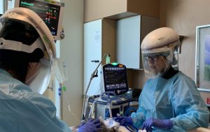
To find out what might be going on, the researchers studied 41 COVID-19 patients hospitalized at University of Utah Hospital in Salt Lake City. Seventeen of these patients were in the ICU, including nine who were on ventilators. They compared blood from these patients with samples taken from healthy individuals who were matched for age and sex.

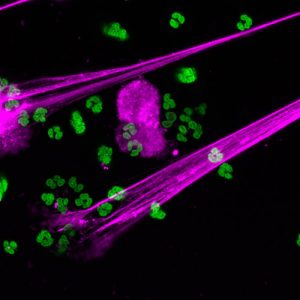
Using differential gene analysis, the researchers found that SARS-CoV-2, the virus that causes COVID-19, appears to trigger genetic changes in platelets. In laboratory studies, they studied platelet aggregation, an important component of blood clot formation, and observed COVID-19 platelets aggregated more readily. They also noted that these changes significantly altered how platelets interacted with the immune system, likely contributing to inflammation of the respiratory tract that may, in turn, result in more severe lung injury.

Surprisingly, Campbell and his colleagues didn’t detect evidence of the virus in the vast majority of platelets, suggesting that it could be promoting the genetic changes within these cells indirectly.

One possible mechanism is inflammation, according to Bhanu Kanth Manne, one of the study’s lead authors and a research associate with the University of Utah Molecular Medicine Program (U2M2). In theory, inflammation caused by COVID-19 could affect megakaryocytes, the cells that produce platelets. As a result, critical genetic alterations are passed down from megakaryocytes to the platelets, which, in turn, make them hyperactive.

In test-tube studies, the researchers found that pre-treating platelets from SARS-CoV-2 infected patients with aspirin did prevent this hyperactivity. These findings suggest aspirin may improve outcomes; however, this will need further study in clinical trials. For now, Campbell warns against using aspirin to treat COVID-19 unless recommended by your physician.

In the meantime, the researchers are beginning to look for other possible treatments.

“There are genetic processes that we can target that would prevent platelets from being changed,” Campbell says. “If we can figure out how COVID-19 is interacting with megakaryocytes or platelets, then we might be able to block that interaction and reduce someone’s risk of developing a blood clot.”

Find original post here.


This study titled, “Platelet Gene Expression and Function in COVID-19 Patients,” was funded by the National Institutes of Health, the University of Utah Health 3i Initiative and the American Heart Foundation.

SARS-CoV-2-like particles very sensitive to temperature

Winter is coming in the northern hemisphere and public health officials are asking how the seasonal shift will impact the spread of SARS-CoV-2, the virus that causes COVID-19?

A new study tested how temperatures and humidity affect the structure of individual SARS-Cov-2 virus-like particles on surfaces. They found that just moderate temperature increases broke down the virus’ structure, while humidity had very little impact. In order to remain infectious, the SARS-Cov-2 membrane needs a specific web of proteins arranged in a particular order. When that structure falls apart, it becomes less infectious. The findings suggest that as temperatures begin to drop, particles on surfaces will remain infectious longer.

This is the first study to analyze the mechanics of the virus on an individual particle level, but the findings agree with large-scale observations of other coronaviruses that appear to infect more people during the winter months.

“You would expect that temperature makes a huge difference, and that’s what we saw. To the point where the packaging of the virus was completely destroyed by even moderate temperature increases,” said Michael Vershinin, assistant professor in the Department of Physics & Astronomy at the University of Utah and co-senior author of the paper. “What’s surprising is how little heat was needed to break them down—surfaces that are warm to the touch, but not hot. The packaging of this virus is very sensitive to temperature.

The paper published online on Nov. 28, 2020, in the journal Biochemical Biophysical Research Communications. The team also published a separate paper on Dec. 14, 2020, in Scientific Reportsdescribing their method for making the individual particle packaging. The virus-like particles are empty shells made from the same lipids and three types of proteins as are on active SARS-Cov-2 viruses, but without the RNA that causes infections. This new method allows scientists to experiment with the virus without risking an outbreak.

The SARS-CoV-2 is commonly spread by exhaling sharply, (e.g. sneezing or coughing), which ejects droplets of tiny aerosols from the lungs. These mucus-y droplets have a high surface to volume ratio and dry out quickly, so both wet and dry virus particles come into contact with a surface or travel directly into a new host. The researchers mimicked these conditions in their experiments.

They tested the virus-like particles on glass surfaces under both dry and humid conditions. Using atomic force microscopy they observed how, if at all, the structures changed. The scientists exposed samples to various temperatures under two conditions: with the particles inside a liquid buffer solution, and with the particles dried out in the open. In both liquid and bare conditions, elevating the temperature to about 93 degrees F for 30 minutes degraded the outer structure. The effect was stronger on the dry particles than on the liquid-protected ones. In contrast, surfaces at about 71 degrees F caused little to no damage, suggesting that particles in room temperature conditions or outside in cooler weather will remain infectious longer.

They saw very little difference under levels of humidity on surfaces, however, the scientists stress that humidity likely does matter when the particles are in the air by affecting how fast the aerosols dry out. The research team is continuing to study the molecular details of virus-like particle degradation.

“When it comes to fighting the spread of this virus, you kind of have to fight every particle individually. And so you need to understand what makes each individual particle degrade,” Vershinin said. “People are also working on vaccines and are trying to understand how the virus is recognized? All of these questions are single-particle questions. And if you understand that, then that enables you to fight a hoard of them.”

Abhimanyu Sharma, Benjamin Preece, Heather Swann, and Saveez Saffarian of the University of Utah and Xiangyu Fan, Richard .J. McKenney and Kassandra M. Ori-McKenney of University of California, Davis were also authors of the Biochem Biophys Res Comms study. Heather Swann,  Abhimanyu Sharma, Benjamin Preece, Abby Peterson, Crystal Eldridge, David M. Belnap and Saveez Saffarian of the University of Utah also co-authored the Scientific Reports study.

Find the original post here.

U studying services for homeless populations during COVID-19

This is adapted from a release by the University of North Texas, available here, and is used with permission.

University of Utah political scientist Jesus Valero is embarking on a study of how the continuum of care for people experiencing homelessness, which includes the social, medical, public health and education sectors, has changed during the COVID-19 pandemic.

Valero and Hee Soun Jang, an associate professor and graduate coordinator for the Department of Public Administration at the University of North Texas, have been researching services for people experiencing homelessness that are run at a local level and often involve multiple agencies known as Continuum of Care.

“Understanding how organizations from the medical, social, and public health systems are working together to address the needs of individuals and families experiencing homelessness during COVID-19 is crucial to improving the effectiveness of community programs,” Valero said.

“Continuum of Care is a premise that is unique to improving a fragmented service system for homelessness,” Jang said. “There are innovative and interesting examples of individual agencies providing successful interventions for homeless populations. But, because agencies do not always coordinate services and information with one another, it is difficult to capture comprehensive knowledge of homeless populations.”

In their new research project, funded by a grant from Systems for Action, a national research program of the Robert Wood Johnson Foundation (RWJF), Valero and Jang are specifically looking at how Continuum of Care networks were affected by COVID-19. They will use previous national studies from 2018 conducted by IBM and RWJF to compare pre-COVID-19 levels of service with current offerings.

“The COVID-19 pandemic has disproportionately affected vulnerable populations and this RWJF-funded project seeks to identify best practices and the conditions that help communities achieve successful collaboration for those experiencing homelessness,” Valero said.

The two-year project will start with case studies in 20 U.S. cities that have both high levels of homelessness and high levels per capita of COVID-19 cases. The second year will consist of a national survey developed from the case studies. Valero and Jang will integrate factors such as racial equity into their research.

“The community has to develop its coordinated approach and aligned mechanism for a fuller, holistic, comprehensive service system that can help these homeless communities,” Jang said. “The people that experience homelessness are in very different stages of their lives. There is no single approach that can fix the problem.”

Findings will be used to understand the effects of the pandemic on Continuum of Care homeless service networks and the effectiveness of the networks in achieving health equity during COVID-19.

“We hope,” Valero said, “that the results of this study will help improve the coordination of cross sector actors in addressing the multidimensional needs of homeless individuals particularly during this public health pandemic.”

See original post here.

Systems for Action is a national research program of the Robert Wood Johnson Foundation that aims to discover and apply new evidence about ways of aligning delivery and financing systems across the medical, public health, and social services sectors that support a Culture of Health.

COVID-19 complication clues

An overactive defense response may lead to increased blood clotting, disease severity and death from COVID-19. A phenomenon called NETosis—in which infection-fighting cells emit a web-like substance to trap invading viruses—is part of an immune response that becomes increasingly hyperactive in people on ventilators and people who die from the disease.

A team led by University of Utah Health and PEEL Therapeutics, in collaboration with Cold Spring Harbor Laboratory and Weill Cornell Medicine, report the findings in a new study published in the journal Blood.

“This study tells us about a potential mechanism for lung injury in COVID-19 that had not previously been recognized as a possible target for treatment,” says Elizabeth Middleton, the study’s first author and a critical care specialist at U of U Health.

The investigation also reports that a naturally occurring protein—originally found in umbilical cord blood—quiets this NET immune response in laboratory experiments, potentially opening new avenues for treatment.

A microscopic image of immune cells emitting a web-like substance in hot pink, with small specks of hot green specks.

Image of infection-fighting cells emitting a web-like substance (hot pink) to trap invading viruses.

It is estimated that up to 10% of people with COVID-19 become critically ill with respiratory distress. Causes of lung damage are a subject of intense investigation, and increasing evidence demonstrates that increased blood clotting may lead to complications caused by the disease.

A research team led by Elizabeth Middleton (pictured), Christian Con Yost and Joshua Schiffman at University of Utah Health has found that a phenomenon called NETosis—in which infection-fighting cells emit a web-like substance to trap invading viruses—is part of an immune response that becomes increasingly hyperactive in people on ventilators and people who die from the disease.

As part of an immune response, white blood cells release web-like Neutrophil Extracellular Traps (NETs) to capture and kill pathogens. While typically beneficial, Yost had previously shown that overactive NETs exacerbate certain illnesses. In conditions such as overwhelming infection, NETs can clog blood vessels and lead to inflammatory tissue damage.

To determine whether NETs could be responsible for complications seen in COVID-19, the team examined plasma from 33 patients, along with tracheal aspirates from the lungs. They found that NET activity correlated with disease severity.

Patients on life support and those who died from COVID-19 had significantly more signs of NET activation than patients who were not as sick or who went on to recover. The NET immune response was lower still in healthy people. NET levels also tracked with a marker for blood-oxygen levels, an independent indicator of disease severity.

Similarly, plasma from sick patients was primed to launch the NET response. When examined in laboratory experiments, plasma from COVID-19 patients triggered white blood cells from healthy patients to shoot out 50 times as many NETs as cells exposed to plasma from otherwise healthy adults.

“This study may tell us that NET levels in the blood could potentially help predict disease severity and mortality in COVID-19,” says Yost. “Additional information is urgently needed in this pandemic regarding how to know which patient will fare better or worse.” Larger studies will need to be done to determine whether NETs could become a biomarker for COVID-19 severity. “Importantly, we think exaggerated NETs could be a cause of morbidity and mortality in COVID-19,” Yost says.

In support of the idea, collaborators at Cold Spring Harbor Laboratory showed that blood vessels in the lungs of deceased COVID-19 patients were dotted with clumps of NET-producing cells and a critical type of blood cell for clotting, the platelets. Another recent study from U of U Health showed that platelets become hyperactive during the disease. Investigations are now underway to determine whether NETs and platelets increase the risk for blood clotting and other clinical manifestations of COVID-19.

Two scientists wearing face visors, blue hospital gowns, and thick blue rubber gloves working in a lab.

A research team led by Elizabeth Middleton (pictured), Christian Con Yost and Joshua Schiffman at University of Utah Health has found that a phenomenon called NETosis—in which infection-fighting cells emit a web-like substance to trap invading viruses—is part of an immune response that becomes increasingly hyperactive in people on ventilators and people who die from the disease.

“In COVID-19, thrombosis is a major cause of death. So, our findings tell us that we should focus on understanding more about NETs’ role in clotting in COVID-19,” Mikala Egeblad, a cancer researcher from Cold Spring Harbor Laboratory, says. “Thrombosis is also a major cause of death in late-stage cancer, where there also can be elevated NETs in the blood. Therefore, I think that what we learn from COVID-19 will help us with other diseases, including cancer.”

Additionally, laboratory experiments showed that a small protein found in the umbilical cord blood of newborn babies, called neonatal NET Inhibitory Factor (nNIF), quiets the hyperactive NET response in white blood cells treated with COVID-19 patient plasma. This peptide is thought to protect babies from harmful inflammation early in life, explains Schiffman, CEO of PEEL Therapeutics. His company is now evaluating whether the protein could become the basis for clinical treatment.

“Newborns babies have a natural therapeutic in their blood to protect against these same inflammatory events that we think could be killing COVID-19 patients,” Schiffman says. “This targeted approach to stopping NETs may be more effective with fewer side effects than some other drugs being tested now in COVID-19 patients that block the entire immune system­­­­.”

Find original post here.

The research was carried out in collaboration with PEEL Therapeutics, Cold Spring Harbor Laboratory, New York Presbyterian Hospital, and Weill Cornell Medicine and published as “Neutrophil Extracellular Traps (NETs) Contribute to Immunothrombosis in COVID-19 Acute Respiratory Distress Syndrome” on June 29, 2020, in Blood.

Support for the work came from the National Institutes of Health, University of Utah Health’s 3i Initiative, Fonds voor Wetenschappelijk Onderzoek Vlaanderen FWO, Animal Cancer Foundation, Soccer for Hope Foundation, Closer to Cure Foundation, PEEL Therapeutics, Inc., William C. and Joyce C. O’Neill Charitable Trust, the Linartz-Meier Family Foundation and the U.S. Department of Veterans Affairs.

First daily surveillance of emerging COVID-19 hot spots

Over the course of the coronavirus epidemic, COVID-19 outbreaks have hit communities across the United States. As clusters of infection shift over time, local officials are forced into a whack-a-mole approach to allocating resources and enacting public health policies. In a new study led by the University of Utah, geographers published the first effort to conduct daily surveillance of emerging COVID-19 hotspots for every county in the contiguous U.S. The researchers hope that timely, localized data will help inform future decisions.

Using innovative space-time statistics, the researchers detected geographic areas where the population had an elevated risk of contracting the virus. They ran the analysis every day using daily COVID-19 case counts from Jan. 22 to June 5, 2020 to establish regional clusters, defined as a collection of disease cases closely grouped in time and space. For the first month, the clusters were very large, especially in the Midwest. Starting on April 25, the clusters become smaller and more numerous, a trend that persists until the end of the study.

The article published online on June 27, 2020, in the journal Spatial and Spatio-temporal Epidemiology. The study builds on the team’s previous work by evaluating the characteristics of each cluster and how the characteristics change as the pandemic unfolds.

Weekly clusters resulting from the prospective space-time scan statistic.

“We applied a clustering method that identifies areas of concern, and also tracks characteristics of the clusters—are they growing or shrinking, what is the population density like, is relative risk increasing or not?” said Alexander Hohl, lead author and assistant professor at the Department of Geography at the U. “We hope this can offer insights into the best strategies for controlling the spread of COVID-19, and to potentially predict future hotspots.”

The research team, including Michael Desjardins of Johns Hopkins Bloomberg School of Public Health’s Spatial Science for Public Health Center and Eric Delmelle and Yu Lan of the University of North Carolina at Charlotte, have created a web application of the clusters that the public can check daily at COVID19scan.net. The app is just a start, Hohl warned. State officials would need to do smaller scale analysis to identify specific locations for intervention.

“The app is meant to show where officials should prioritize efforts—it’s not telling you where you will or will not contract the virus,” Hohl said. “I see this more as an inspiration, rather than a concrete tool, to guide authorities to prevent or respond to outbreaks. It also gives the public a way to see what we’re doing.”

The researchers used daily case counts reported in the COVID-19 Data Repository from the Center for Systems Science and Engineering at Johns Hopkins University, which lists cases at the county level in the contiguous U.S. They used the U.S. Census website’s 2018 five-year population estimates within each county.

To establish the clusters, they ran a space-time scan statistic that takes into account the observed number of cases and the underlying population within a given geographic area and timespan. Using SatScan, a widely used software, they identified areas of significantly elevated risk of COVID-19. Due to the large variation between counties, evaluating risk is tricky. Rural areas and small, single counties may not have large populations, therefore just a handful of cases would make risk go up significantly.

This study is the third of the research group’s iteration using the statistical method for detecting and monitoring COVID-19 clusters in the U.S. Back in May, the group published their first geographical study to use the tracking method, which was also of the first paper published by geographers analyzing COVID-19. In June, they published an update.

“May seems like an eternity ago because the pandemic is changing so rapidly,” Hohl said. “We continue to get feedback from the research community and are always trying to make the method better. This is just one method to zero in on communities that are at risk.”

A big limitation of the analysis is the data itself. COVID-19 reporting is different for each state. There’s no uniform way that information flows from the labs that confirm the diagnoses, to the state health agencies to the COVID-19 Data Repository from the Center for Systems Science and Engineering at Johns Hopkins University, where the study gets its data. Also, the testing efforts are quite different between states, and the team is working to adjust the number of observed cases to reflect a state’s efforts. Hohl is also working with other U researchers to look at the relationship between social media and COVID-19 to predict the future trajectory of outbreaks.

“We’ve been working on this since COVID-19 first started and the field is moving incredibly fast,” said Hohl. “It’s so important to get the word out and react to what else is being published so we can take the next step in the project.”

Find the original post here.

The overlooked casualties of COVID-19

The daily toll of COVID-19, as measured by new cases and the growing number of deaths, overlooks a shadowy set of casualties: The rising risk of mental health problems among health care professionals working on the frontlines of the pandemic.

A new study, led by University of Utah Health scientists, suggests more than half of doctors, nurses and emergency responders involved in COVID-19 care could be at risk for one or more mental health problems, including acute traumatic stress, depression, anxiety, problematic alcohol use and insomnia. The researchers found that the risk of these mental health conditions was comparable to rates observed during natural disasters, such as 9/11 and Hurricane Katrina.

Headshot of a male with brown hair, black suit jacket, blue tie, striped collared shirt, against a white background.

Andrew J. Smith, director of the U of U Health Occupational Trauma Program at the Huntsman Mental Health Institute.

“What health care workers are experiencing is akin to domestic combat,” says Andrew J. Smith, director of the U of U Health Occupational Trauma Program at the Huntsman Mental Health Institute and the study’s corresponding author. “Although the majority of health care professionals and emergency responders aren’t necessarily going to develop PTSD, they are working under severe duress, day after day, with a lot of unknowns. Some will be susceptible to a host of stress-related mental health consequences. By studying both resilient and pathological trajectories, we can build a scaffold for constructing evidence-based interventions for both individuals and public health systems.”

The study appears in the Journal of Psychiatric Research. In addition to U of U Health scientists, contributors include researchers from the University of Arkansas for Medical Sciences; University of Colorado, Colorado Springs; Central Arkansas VA Health Care System; Salt Lake City VA Healthcare System; and the National Institute for Human Resilience.

The researchers surveyed 571 health care workers, including 473 emergency responders (firefighters, police, EMTs) and 98 hospital staff (doctors, nurses), in the Mountain West between April 1 and May 7, 2020. Overall, 56% of the respondents screened positive for at least one mental health disorder. The prevalence for each specific disorder ranged from 15% to 30% of the respondents, with problematic alcohol use, insomnia and depression topping the list.

“Frontline providers are exhausted, not only from the impact of the pandemic itself, but also in terms of coping day to day,” says Charles C. Benight, co-author of the study and a professor of psychology at the University of Colorado, Colorado Springs. “They’re trying to make sure that their families are safe [and] they’re frustrated over not having the pandemic under control. Those things create the sort of burnout, trauma and stress that lead to the mental health challenges we’re seeing among these caregivers.”

In particular, the scientists found that health care workers who were exposed to the virus or who were at greater risk of infection because they were immunocompromised had a significantly increased risk of acute traumatic stress, anxiety and depression. The researchers suggest that identifying these individuals and offering them alternative roles could reduce anxiety, fear and the sense of helplessness associated with becoming infected.

Alcohol abuse was another area of concern. About 36% of health care workers reported risky alcohol usage. In comparison, estimates suggest that less than 21% of physicians and 23% of emergency responders abuse alcohol in typical circumstances. Caregivers who provided direct patient care or who were in supervisory positions were at greatest risk, according to the researchers. They say offering these workers preventative education and alcohol abuse treatment is vital.

Surprisingly, health care workers in this study felt less anxious as they treated more COVID-19 cases.

“As these health care professionals heard about cases elsewhere before COVID-19 was detected in their communities, their anxiety levels likely rose in anticipation of having to confront the disease,” Smith says. “But when the disease started trickling in where they were, perhaps it grounded them back to their mission and purpose. They saw the need and they were in there fighting and working hard to make a difference with their knowledge and skills, even at risk to themselves.”

Among the study’s limitations are its small sample size. It was also conducted early in the pandemic in a region that wasn’t as affected by the disease as other areas with higher infection and death rates.

Moving forward, the researchers are in the final stages of a similar but larger study conducted in late 2020 that they hope will build on these findings.

“This pandemic, as horrific as it is, offers us the opportunity to better understand the extraordinary mental stress and strains that health care providers are dealing with right now,” Smith says. “With that understanding, perhaps we can develop ways to mitigate these problems and help health care workers and emergency responders better cope with these sorts of challenges in the future.”

Find original post here.


In addition to Smith, Hannah M. Wright, Tiffany M. Love and Scott A. Langenecker of University of Utah Health contributed to this study. The study, “Pandemic-related mental health risk among front line personnel,” was published in the Journal of Psychiatric Research.

U designs innovative respirator system

In response to the overwhelming demand for Personal Protective Equipment (PPE) due to COVID-19, the Center for Medical Innovation (CMI) at University of Utah Health has designed an enclosed Powered Air Purifying Respirator (PAPR) system to provide health care workers safe and reusable PPE when working with COVID-19 patients.


Designed to fully enclose the face of the user, a PAPR system consists of a hood or helmet and a filtered respirator to provide those wearing the system a constant flow of clean air. This positive pressure prevents entry of unfiltered air and protects the health worker from inhaling aerosolized COVID-19 particles.

However, due to the specialized nature of the equipment, hospitals typically have a limited quantity of the systems on-hand at any given time.

“PAPR systems provide excellent protection and can drastically reduce the consumption of single-use PPE, such as common N95 respirators,” explains Bryan McRae, interim co-director at CMI. “Unfortunately, PAPRs have now been unavailable for standard suppliers for more than a month. The CMI team and our University of Utah Health colleagues have been nimble and innovative in developing a solution to bridge the gap while traditional PPE sources remain uncertain.”

By combining readily available components such as mobile battery packs, portable fans and replaceable medical grade filters with several 3-D printed adapters, the assembled PAPR system designed by CMI expands the options to protect health workers treating COVID-19 patients.

The PAPR system on a table. The components are a plastic face shield surrounded by a white material that looks like a shower cap, connected to a long tube that plugs into a portable air filtration system.

The PAPR system is made by combining readily available components such as mobile battery packs, portable fans, and replaceable medical grade filters with several 3D printed adapters.

Using a standardized rating system known as “Fit Factor,” PAPR systems typically rate somewhere between 200 and 1000 on the quantitative fit testing scale. This means the system reduces the concentration of 0.3 micron aerosolized particles inside the system by 200 to 1000 times when compared to the air outside the hood. The PAPR system developed by the CMI was assessed by the Rocky Mountain Center for Occupational and Environmental Health at the University of Utah and offers a Fit Factor of 400 or better.

For comparison, on the OSHA scale known as an Assigned Protection Factor (APF), this PAPR offers an APF between 25 and 400, providing superior protection when compared to the common N95 respirator masks which typically only provide an APF of 10.

As the University of Utah Hospital braces for a potential surge of COVID-19 patients, all options to expand the supply of PPE are being explored. This includes retrofitting older pieces of still-viable equipment to the newer PAPR systems. Because of the customized 3-D printed adapter used to connect the respirator to the helmet, CMI’s PAPR system can also connect to older models of PAPR helmets still in-stock, enabling hundreds of previously unusable helmets to be worn safely and comfortably by health care workers at University Hospital.

Integrating feedback directly from those on the frontlines of COVID-19 management into the final PAPR system design, production has already begun to manufacture several hundred units as quickly as possible. Utilizing 3-D printing equipment on campus, including over 30 printers in production at the Marriott Library, Eccles Health Sciences Library and the College of Engineering, many of the PAPR systems will be ready for implementation within the week. Additional manufacturing support is being generously provided by O.C. Tanner and L3 Technologies.

“We are especially grateful for the expertise and insight from our university and industry partners,” said Bernhard Fassl, interim co-director of CMI. “As we further develop solutions for health workers during the COVID-19 outbreak, we will continue to rely on our community partners to help us implement these projects.”

Later this week, CMI will be releasing design specifications for the PAPR system to other health care groups and the public. This includes Indian Health Services and the Navajo Nation, as well as CMI’s global health partners in India, Kenya and Nepal, to provide guidance on assembly and use in resource-limited settings.

For more information about the PAPR system and the Center for Medical Innovation’s response to the COVID-19 outbreak, please click here.

Find original post here.

Portable, reusable test for COVID-19

“Testing, testing, testing.” It’s a mantra that health officials have been constantly promoting because screening people for COVID-19 is the best way to contain its spread. In the U.S., however, that crucial necessity has been hampered due to a lack of supplies.

But University of Utah electrical and computer engineering professor Massood Tabib-Azar has received a $200,000 National Science Foundation Rapid Response Research (RAPID) grant to develop a portable, reusable coronavirus sensor that people can always carry with them. The sensor, about the size of a quarter, works with a cellphone and can detect COVID-19 in just 60 seconds.

“It can be made to be a standalone device, but it can also be connected to a cellphone,” says Tabib-Azar. “Once you have it connected either wirelessly or directly, you can use the cellphone software and processor to give a warning if you have the virus.”

Health officials say the U.S. needs to conduct at least five million COVID-19 tests per day to effectively understand and contain the spread of the virus. But at most, 319,000 per day have been given, according to The COVID Tracking Project, mostly due to a lack of testing supplies such as swabs and reagents. Typically, a 6-inch swab is inserted through the nose to the back of the cavity for 15 seconds to obtain a sample that is sent to a lab for analysis. Most tests take between four to seven days for the results.

A headshot of an engineer standing in front of technological equipment, in a brown jacket, red tie, blue shirt, wearing glasses.

University of Utah electrical and computer engineering professor Massood Tabib-Azar.

Tabib-Azar’s technology, which was profiled in two papers published last month in IEEE Sensors Journal, involves just a drop of saliva and can produce results in a minute. It is based on a sensor Tabib-Azar first began developing for the NSF about a year ago to detect the Zika virus. He is now converting the same technology to work with COVID-19.

The sensor would use single-strand DNA called aptamers in the sensor that would attach to the proteins in the COVID-19 virus molecule if it is present. A person would plug the small sensor into the cellphone’s power jack and launch an app made for the device. To test for the presence of the virus, the user would place a drop of saliva on the sensor, and the results would appear on the phone. It is designed to also test for the virus on the surface of something, like a table or desk, by brushing a swab on the surface and then on the sensor. And it might be able to detect the presence of COVID-19 in floating microscopic particles in the air in enclosed spaces such as an elevator (while the virus is currently considered not airborne, studies are being conducted to determine if minute particles of the virus can hang in floating droplets in the air.).

If the virus is present, the DNA strands in the sensor would bind to the virus’ proteins and electrical resistance is measured in the device, signaling a positive result.

Tabib-Azar says the sensor would include an array of tiny devices inside it, each with a DNA strand that looks for a different protein. A specific combination of proteins would be unique to just COVID-19.

“By increasing the number of devices and single-strand DNA, we can increase the sensor’s accuracy and reduce the false positives and false negatives,” he says.

The sensor is designed to be reusable because it can destroy the previous sample on it by producing a small electrical current that could heat up and remove or disintegrate the virus. Tabib-Azar says the entire process would use little battery power from the cellphone.

Another possible method would involve putting the saliva sample on disposable sheets that are placed on top of the sensor like a sticky note. This would decrease cross-contamination on the sensor and eliminate the need to heat up and destroy the sample afterward.

The device also can be designed to upload the results to a central server that maps out positive results in an area, giving researchers a clearer and more accurate picture of where hotspots are with big outbreaks of the virus.

Because Tabib-Azar has already developed the technology—and a prototype—to detect the Zika virus, he said he could have a prototype of the new COVID-19 sensor for clinical trials in two to three months.